Patients need a cleansing preparation of their bowel
prior to the test. The actual VC procedure will begin by having a small
flexible rubber tube placed in the rectum, so that air can be introduced. A
helical CT scan is then performed while the patient lies comfortably on the
back (supine) and then on the stomach (prone). The total time required for
the study is around 15 minutes. Because sedation is not required, patients
are free to leave the CT suite immediately without the need for observation
or recovery. Patients can resume normal activities immediately after the
procedure and can eat, work or drive without a delay. When air is introduced
in the colon, some patients experience minimal temporary abdominal cramping
or "gas pains."
ACCURACY
Research has shown that Virtual Colonoscopy is better
able to see polyps than Barium Enema and is nearly as accurate as
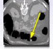
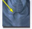
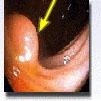
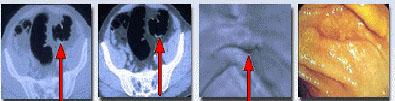
Conventional Colonoscopy. Fig. 1 shows a polyp and Fig. 2
a flat cancer seen on 3 techniques: 2D axial CT (Fig. 1a, 2a-b), 3D
VC (Fig. 1b, 2c), and conventional colonoscopy (Fig. 1c, 2d).
Most patients report that the Virtual Colonoscopy technique is more
comfortable than either Barium Enema or Conventional Colonoscopy. Studies
suggest a very high sensitivity and specificity (96%) for the detection of
polyps 1 cm or greater. Such polyps have significant malignant potential.
Sensitivity for polyps less than 1 cm is significantly less. Although
controversy exists as to the definition of a "significant" polyp with regard
to size, polyps < 1 cm in size have a low probability of malignancy and the
likelihood of any single lesion progressing to cancer is also small.
INDICATIONS
Indications for VC include screening for polyps,
incomplete or failed colonoscopy, and preoperative assessment of the colon
proximal to an occlusive cancer (defined as a tumor that cannot be traversed
endoscopically). Virtual colonoscopy excels in the evaluation of the
ascending colon, particularly the cecum due to the degree of distension
achievable and the typical lack of spasm or muscular hypertrophy, which is
seen in the sigmoid colon. It is therefore a useful complement to an
incomplete colonoscopy. Any questionable abnormality will warrant a
conventional colonoscopy procedure, thus resulting in GI referrals.
VC may become a screening tool for detecting colorectal
neoplasia, potentially supplanting Conventional Colonoscopy as a tool for
detecting lesions. Increased screening, with increased detection, should
decrease the incidence of colorectal cancer, as premalignant growths can be
found at an earlier stage. About 5% of colon cancers (flat cancers) arise
from normal mucosa and not from polyps and are difficult to identify on
conventional and virtual colonoscopy, while axial helical CT images improve
detection of such cancers.
HealthScan Program
AIC has been offering for some time a screening
total-body scan program called HealthScan,
which includes a screening helical CT of the torso, helical CT coronary
calcium scoring, and more optional 3D virtual colonoscopy.

 1.
The only community-based, private-practice, physician-operated
imaging facility in the Antelope Valley, just like any other private
practice medical office. Not belonging to any hospital or outside
imaging network. This means more personal and caring service.
1.
The only community-based, private-practice, physician-operated
imaging facility in the Antelope Valley, just like any other private
practice medical office. Not belonging to any hospital or outside
imaging network. This means more personal and caring service.