Q. What is Coronary Calcification Scoring?
Coronary calcification scoring or cardiac scoring
is a CT technique to determine the amount of calcium build up in the
coronary arteries. Coronary artery calcification is a specific marker
for coronary atherosclerosis. The amount of calcification correlates
with severity of coronary atherosclerosis and the probability of
obstructive disease.
Q. How is it performed?

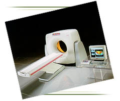
The scan is performed on an ultrafast CT (either
helical or electron beam CT with similar accuracy) in one breathhold. At
AIC a 16-slice ultrafast multi-slice, multi-detector helical CT is used,
and the whole procedure takes just a few minutes.
Q. What happens after the scan?
The data are processed via a special cardiac
scoring software package. A radiologist then evaluates the images and
puts region of interests (ROI's) on the calcified coronary arteries.
At the end, individual scores for four arteries (left main, LAD,
circumflex, and right coronary) and a total score are calculated. The
total score falls under one of the following categories:
0-1: NO CALCIFICATION (extremely low likelihood for
obstructive coronary disease);
1-10: MINIMAL CALCIFICATION;
11-100: SMALL AMOUNT CALCIFICATION;
101-400: MODERATE CALCIFICATION;
>400: LARGE AMOUNT CALCIFICATION (high likelihood for
extensive coronary atherosclerosis).
Q. What's the accuracy of the test?
It has a nearly 100% sensitivity for calcifications
and nearly 100% negative predictive value for future coronary events.
The positive predictive value ranges from 50 to 80. A zero or very low
score implies virtually no coronary obstructive disease with the
exception occurring in young patients who smoke (soft plaques). A high
score indicates a significant plaque burden and risk for future
cardiovascular event. It should be understood that calcification is
not site specific for stenosis but rather indicates the extent of
atherosclerosis in the coronary arteries overall. The score may be
used as benchmark to measure subsequent disease development or assess
preventive programs.
Q. Who should get this test?
Individuals who have any of the following: history
of smoking, diabetes, hypertension, hypercholesterolemia, family
history of coronary artery disease, obesity, sedentary lifestyle, high
level of stress, atypical chest pain, asymptomatic males over 45 and
females over 55 years of age.
 |
 |
 |
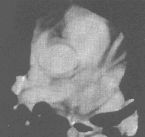
| No calcification |
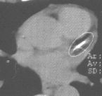
Calcification in LAD coronary artery |
Coronary artery disease (CAD) is the leading
cause of death in the United States. Each year 30-50% of the 1.5 million
American men and women who have a heart attack (myocardial infarct) die
as a result. Most of these occur in people who've had no previous
symptoms or warning. Men are at greater risk for heart attack at certain
times of their lives, but overall men and women die at equal rates from
CAD.
Coronary artery calcification scoring is a
non-invasive CT scan of the heart performed on our 16-slice
Helical CT. The
scan detects and quantifies calcified atherosclerotic plaque in the
coronary arteries. A score is computed based on the amount of
calcification detected. Your score is an accurate predictor of the
degree of narrowing of the coronary arteries and the likelihood of a
future coronary event (heart attack). The radiologist, an imaging
specialist, will interpret the scans and send a report to you and your
physician.
The American Heart Association has identified the following risk
factors:
- Men over age 45
- Women over age 55
- Elevated LDL cholesterol
- Low HDL cholesterol
- Family history of coronary artery disease
- Smoking
- Obesity
- Sedentary life style
- High blood pressure
- Diabetes
If you are a male over 45 or a female over 55 and have one or more of
these risk factors,
CORONARY CALCIFICATION SCORING
WILL BE USEFUL TO YOU!
500,000 Americans die from coronary artery disease
(heart attack) yearly. Most have no warning prior to their death! Early
detection of calcified atherosclerotic plaque can prompt preventive
action to minimize risk of heart attack or direct you to seek medical
evaluation for further testing. Coronary atherosclerosis can be slowed,
stopped, and possibly reversed before artery blockage results in heart
muscle damage or death.
Remember, in cardiac disease, PREVENTION could mean
everything!
Coronary Artery Calcification Scoring has clinical value:
- To determine if patients with chronic atypical chest pain have
coronary atherosclerosis.
- To screen asymptomatic patients in order to stratify their risk
of coronary disease and future cardiac events.
- To exclude the presence of coronary artery disease in women over
60 years of age.
- To determine if patients with equivocal stress or thallium test
have coronary artery plaque before proceeding to coronary
angiography.
- To identify postmenopausal women with low risk for coronary
disease who are therefore less likely to benefit from
cardioprotective effects of hormone replacement therapy.
- To determine if a dilated cardiomyopathy is secondary to
coronary artery disease or not.
- To simplify the pre-operative cardiac clearance of women over
age 60.
- To follow the progression of coronary artery plaque
non-invasively.
Read selected journal references for Coronary Artery Calcification
Scoring.
Q & A regarding AIC's Ultrafast, multidetector
Helical CT
Q. Can you tell me about the new helical CT at AIC?
A. Certainly. It is a 16-slice CT. The new helical CT has
replaced our old dual-slice CT. It is a multi-slice (multi-detector) CT
capable of simultaneously scanning 16 slices evry 0.4 second (or 40
slices per second), thus increasing the speed of CT scanning by at least
a factor of 40 or more. It is the fastest CT in the Antelope Valley
area.
Q. What does ultrafast CT allow you to do?
A. Here's a summary (* denotes AIC exclusive):
- Fast high-resolution (1-2 mm) routine imaging of the
neck, chest, abdomen, and pelvis.
- *Fast high-res (0.5 mm) imaging of the IAC's/Temporal bones.
- *Fast ultra-high-resolution (0.5 mm) imaging of the bones for 4D
isotropic reconstruction.
- 3D and 4D CT Angiography (CTA): aorta, pulmonary
arteries, runoffs, brain.
- Coronary artery calcification scoring (excellent
non-invasive screening test).
- Virtual endoscopy (colonoscopy, bronchoscopy, and
endovascular angioscopy with CT!)
Q. What does coronary calcification scoring tell you?
A. Coronary scoring is a safe, noninvasive and fast
screening CT technique that scans the heart in a few seconds and gives a
score based on the amount of calcium build up in the coronary arteries.
The score is a good predictor of future coronary events. For instance,
it has a 100% negative predictive value!
Q. Can you tell me more about the new workstation at AIC?
A. Certainly. The new workstation is a state-of-the-art
silicon graphics workstation linked to all our modalities including
Open MRI, high-field MRI, Helical CT, and Nuclear med (SPECT/PET).
It is a powerful and sophisticated computer capable of amazing
multiplanar and 4D reconstruction, fusion of images from
different modalities, 4D virtual endoscopy, dental scan, 3D/4D CT
Angiography (CTA), just to name a few. It is simply technology at
its best.
Q. What is virtual endoscopy?
A. This noninvasive technique is one of the hottest areas
in CT today that allows 4D visualization of hollow organs (colon,
bronchus, etc.) similar to video endoscopy. This is only available at
AIC.
Q. I have not heard of image Fusion. Can you explain?
A. Of course. Image fusion is a sophisticated software
utilized by our workstation that allows fusion of images from different
modalities (e.g., CT, MRI, SPECT, PET) or fusion of images from the same
modality at different times (to evaluate for growth). For example, a
physiologic/metabolic SPECT or PET image can be combined with an
anatomic CT or MRI image to provide an anatomico-physiologic
image.
 1.
The only community-based, private-practice, physician-operated
imaging facility in the Antelope Valley, just like any other private
practice medical office. Not belonging to any hospital or outside
imaging network. This means more personal and caring service.
1.
The only community-based, private-practice, physician-operated
imaging facility in the Antelope Valley, just like any other private
practice medical office. Not belonging to any hospital or outside
imaging network. This means more personal and caring service.