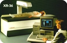
Advanced Imaging Center offers one of the world's
most sophisticated DEXA scanners.
DEXA (stands for Dual Energy X-ray Absorptiometry) is
the safest and most accurate method to measure Bone Mineral Density, an
overall measure of how much calcium there is in the bones.
A low level of BMD below a certain threshold (below 2
standard deviations from peak bone mass) indicates osteoporosis.
DEXA provides low cost, state-of-the-art Bone
Densitometry with insignificant radiation, unlike CT or nuclear bone
densitometry.
Osteoporosis is a common disorder affecting a
large number of adults. In the United States more than 25 million people
are afflicted, particularly women. Many suffer disabling fractures of
the spine, which is the most common site of involvement. Osteoporosis is
believed to be responsible for about 1.3 million fractures annually
including more than 500,000 spine, 250,000 hip, and 240,000 wrist
fractures.
Up to 30% of elderly people with hip fractures die
within 6 months of their injury. The difference in sex distribution in
osteoporosis is especially significant as women who are 65 years of age
or older represent the fastest growing segment of the population in the
United States. The worldwide increase in life expectancy will most
likely result in an accompanying rise in the prevalence of osteoporotic
fractures of all kinds over the next decades.
During the past decades, osteoporosis, called the
"silent epidemic," has gained increased attention. The involvement
chiefly of women and the insidious loss of bone manifested primarily as
"crush fractures" of the spine, hip and wrist are widely known facts.
Public awareness of this disorder also has been heightened by the
resulting increase in health care expenditure that is currently
estimated to be in excess of 7 billion dollars.
Indications for DEXA:
- Patients receiving long term glucocorticoid therapy.
- Patients with primary asymptomatic hyperparathyroidism
- Patients at high risk for osteoporosis such as amenorrhea,
anorexia nervosa or alcoholism
- Patients with atraumatic fractures, disuse atrophy, and similar
conditions
- Assessment of early postmenopausal bone loss as an indication to
initiate estrogen replacement therapy
- Diagnosis of osteoporosis suspected from radiographic findings
or from clinical risk factors
- Serial assessment of bone density, i.e., during treatment for
osteoporosis or in anticipation of rapid bone loss
A variety of metabolic disorders such as
hyperparathyroidism, renal insufficiency, Cushing's syndrome, and
amenorrhea in premenopausal women as well as chronic immobilization and
chronic steroid or thyroid therapy are known to influence calcium
metabolism and may affect the skeleton adversely. In these cases of
secondary osteoporosis, bone density measurements are of particular
importance because they may prompt therapeutic decisions such as
reduction in medication or surgery.
Bone turnover increases significantly at menopause
with a greater increase in bone resorption than bone formation resulting
in accelerated loss of bone. One-third to one-half of bone loss in women
may be attributable to the loss of ovarian function. Several studies
have established the bone mass-preserving effect of estrogen therapy; if
begun soon after menopause it reduces the subsequent rate of vertebral
fractures by 50%. The benefits derived from estrogen therapy clearly
seem to outweigh its adverse effects. However, it is unacceptable for
many women and the level of bone mineral density at menopause and the
magnitude of subsequent loss are important considerations in assessing
the future risk of fracture and a decision to begin prophylaxis can be
based on such considerations.
Serial measurement of bone density is accurate,
providing guidance for clinical treatment. Measurements every 1-2 years
are useful depending on the disease process. Studies have shown large
annual loss of bone density from sites rich in trabecular bone in
patients receiving high dose steroids. Similarly, large annual gains of
bone have been observed in osteoporotic patients receiving treatment
with a variety of therapeutic agents such as calcitonin, fluoride,
biphosphonates, or parathyroid hormone. Considering the marked effect of
some types of intervention, the magnitude of postmenopausal loss of
bone, and the continued improvements in measurement precision, serial
DEXA bone densitometry is valuable in determining therapeutic efficacy.
ScanHealth |
Open MRI |
High-field MRI | MR Angiography |
Helical CT |
CT Angiography |
Calcium Scoring
4D CT Reconstruction |
Dental Scan | 4D
Ultrasound Nuclear Medicine |
PET Scan | DEXA Bone Density |
X-ray

 1.
The only community-based, private-practice, physician-operated
imaging facility in the Antelope Valley, just like any other private
practice medical office. Not belonging to any hospital or outside
imaging network. This means more personal and caring service.
1.
The only community-based, private-practice, physician-operated
imaging facility in the Antelope Valley, just like any other private
practice medical office. Not belonging to any hospital or outside
imaging network. This means more personal and caring service.