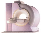
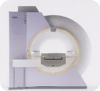
Advanced Imaging Center also provides the first
short-bore, wide flared high-field (1.5 Tesla) MRI in the valley, the
first one of its kind in Southern California, which is the most advanced
MRI system in the world (top right). Same field strength as the closed
MRI's (i.e., 1.5 Tesla) but with open features, this Siemens Symphony
MRI with high-performance gradients is unmatched in the area.
It is capable of performing the most complex MRI
studies, including tasks not available anywhere else in the area (e.g.,
Perfusion/Diffusion study of the brain for early stroke detection;
Breast MRI for implants and cancer; CSF Flow studies; Contrast-enhanced
peripheral MRA with power injector; MRCP [MR version of ERCP]; MR
Myelography; ultra fast single-breath hold abdominal scanning; MR
neurogram; high-performance gradients, thus ultra fast (sub second) and
ultra high-res (1024 matrix); just to name a few). AIC is the only
facility in the Antelope Valley that is MRI certified (accredited by the
American College of Radiology).
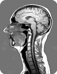
 MRI,
magnetic resonance imaging, is a method used by physicians to visualize
internal organs of the human body and obtain diagnostic information.
These images are produced without the use of radiation. MRI is a
noninvasive procedure and there are no known side or after effects. The
procedure is painless. You won't see or feel anything during the exam. A
faint knocking sound will be heard which is the imaging process in
operation.
MRI,
magnetic resonance imaging, is a method used by physicians to visualize
internal organs of the human body and obtain diagnostic information.
These images are produced without the use of radiation. MRI is a
noninvasive procedure and there are no known side or after effects. The
procedure is painless. You won't see or feel anything during the exam. A
faint knocking sound will be heard which is the imaging process in
operation.
MRI utilizes the physical properties of magnetic
fields, radio waves, and computers to generate images of the body in any
plane. The technique is an established, accurate diagnostic tool. It can
quickly and noninvasively provide accurate diagnosis for your physician,
which in some situations will reduce the need for exploratory surgery or
other diagnostic procedures, which may have greater risk.
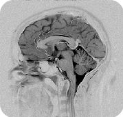
The benefits of magnetic resonance imaging are vast
and new applications are continually being developed through ongoing
research. MRI imaging is used for virtually all parts of the body. It is
the primary imaging modality for evaluation of diseases of the brain and
spine. It is effective in depicting abnormalities of the eye, paranasal
sinuses, throat, salivary glands, and the thyroid.
MRI is the method of choice for imaging of the
musculoskeletal system and is widely used for evaluation of the
shoulder, elbow, wrist, hip, knee, and ankle. It can also accurately
depict abnormalities within the bone marrow. It has many applications in
the cardiovascular system. The display of blood vessels known as MR
angiography is an accurate, noninvasive means of obtaining information
about arteries of the head, neck, and body.
It is also effective in demonstrating congenital
cardiac abnormalities. MRI is also important in evaluation of the organs
of the abdomen and pelvis including the liver, pancreas, kidneys,
ovaries, uterus, and prostate.
Q&A regarding Advanced Imaging Center's New
OPEN High-Field (1.5 Tesla) MRI, the first of its kind in Southern
California
Question:
How Many MRI systems does AIC have?
 Answer:
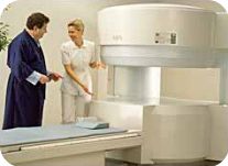
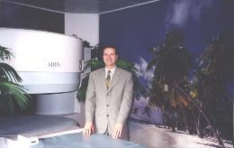
We have two magnets: the OPEN-AIR (Open-Sided)
Hitachi AIRIS (right) and the
1.5 Tesla Siemens Magnetom Symphony
(below).
Question:
Is it true that the new system was the first one in Southern
California?
Answer:
Yes, and only the second one in the state. It is the
next-generation MRI system and the most sophisticated in the world.
Question:
What is the field strength and some of its unique features?
Answer:
It is a high-field 1.5 Tesla system (similar to the "closed
tunnel" systems). But it has a short-bore with patient-friendly
OPEN features (somewhat donut shaped like a CT but longer, as
shown in the top right).
Question:
I've heard that it has high power ... how so?
Answer:
It has quantum (high-performance) gradients that allow for ultra
fast and ultra high resolution (1024 matrix) scanning, not
available elsewhere in the Antelope Valley area. For example, it is
capable of obtaining a single brain slice in less than a second, ideal
for trauma or uncooperative patients.
Question:
What can you do in the NEURO/BRAIN areas with this magnet
not possible on other magnets in the area?
Answer:
Here are some of this system's unique features in the CNS arena:
- Functional imaging such as perfusion/diffusion
imaging capable of diagnosing acute stroke immediately
(crucial for starting early thrombolytic therapy) and
differentiating between chronic and acute strokes and white matter
lesions.
- With its revolutionary CSF Quantitative Flow Analysis
package, the CSF velocity in the brain may be measured (totally
non-invasive). Applications: (1) selection of shunt-responsive
NPH (Normal Pressure Hydrocephalus) patients; (2) accurate
diagnosis of VP shunt patency/shunt malfunction.
- MR neurogram.
Question:
What exactly is an MR neurogram?
Answer:
It is somewhat similar to an MR Angiogram except, instead of
imaging the vessels, the nerves are imaged. Applications: brachial
plexus, lumbar plexus, cranial nerves, or any other nerve for that
matter (ulnar and radial nerves in the wrist and elbow, sciatic
nerve in pyriformis entrapment).
Question:
How about in the head and neck (ENT) area?
Answer:
Head and neck requires exquisite details. Its ultrafast and ultra
high resolution (1024 matrix as opposed to current 256 matrices) allow
exquisite imaging of the head and neck anatomy, such as depiction of
individual 7th and 8th nerves in the internal auditory canals as well
as other cranial nerve anatomy (MR neurogram).
Question:
How about in the ORTHOPEDIC area?
Answer:
It obviously produces exquisite imaging of the joints and spine.
This becomes more apparent in small joints such as the ankle, elbow
and wrist (ultra high resolution of TFC, interosseus ligaments, etc.).
Question:
What about the fingers and toes?
Answer:
Funny you should ask. We actually purchased a digit coil that's
only a couple of inches long, ideal for imaging the digits (e.g., an
MRI image of the finger would now look as big as a knee because of the
ultra small coil!). With this tiny coil, MRI becomes more accurate
than bone scan for accurate and early diagnosis of osteomyelitis.
Question:
What about in the SPINE?
Answer:
Here're some of the advantages of the new system:
- First this revolutionary system allows connection of multiple
coils so that the entire spine (and head) can be done without moving
the patient or changing coils, saving time and adding tremendous
comfort for the patient
- Its 1024 matrix imaging in the spine provides unsurpassed
resolution in the cord and spinal nerve roots.
- MR Myelogram (totally noninvasive) can be done in a minute or
so.
- MR Neurogram as explained above (e.g., for lumbar and brachial
plexus, sciatic nerve, etc.)
Question:
How about UROLOGIC applications?
Answer:
Here's a limited list:
- MRU (MR version of urography similar to IVP and retrograde
pyelogram).
- Prostate cancer detection and staging; Renal cancer and staging
(e.g., invasion of renal veins and IVC)
- Dialysis shunt evaluation using Advanced MR Angio package.
Question:
Tell me more about its MR Angio/Vascular capabilities.
Answer:
It has the most sophisticated MRA package available anywhere such
as:
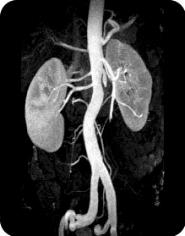
- 20-second Gadolinium-enhanced MRA of the carotids utilizing an
MR-compatible power injector;
- 30-second Gadolinium-enhanced body MRA (pulmonary arteries,
renal arteries, aorta, mesenteric angio;
- 30-second Gadolinium-enhanced peripheral MRA (run-offs utilizing
a new automatic table-moving feature and a power injector with
accuracy similar to or better than X-ray angio).
- Exquisite ultra-resolution MRA of the circle of Willis and the
rest of the brain using Advanced MRA..
Question:
In the GI area, I have heard of MRCP. What is it exactly?
Answer:
MRCP (MR CholangioPancreatography) is the MR version of ERCP, but
is totally non-invasive and takes just a few minutes to do it. Its
accuracy is similar to ERCP in terms of diagnosis of biliary /
gallbladder disease and causes for biliary obstruction (but is
obviously not therapeutic).
Question:
Are there any OB/GYN or Female Imaging applications with this MRI?
Answer:
Definitely. Here's a limited list: Uterine anomalies, detection of
focal fibroids (to avoid total hysterectomy), endometrial disease,
endometriosis, adenomyosis, placenta accreta and other pathologies,
certain intrauterine fetal anomalies, adnexal masses, breast masses
and implants (require a dedicated breast coil).
Question:
Do you have a dedicated bilateral breast coil?
Answer:
Yes, the only one in the Antelope Valley to our best knowledge.
Question:
What are the clinical applications of Breast MRI?
Answer:
Three main applications:
- Detection of implant rupture (intra or extracapsular) with high
accuracy;
- Screening exam in women with dense breasts (where mammography is
limited);
- Differentiating between a benign and malignant breast mass
(utilizing a new Gadolinium-uptake quantitative analysis program).
Question:
How can MRI differentiate between benign and malignant breast
masses?
Answer:
Cancers tend to take up Gadolinium contrast much faster than benign
fibroadenomas. Uptake graphs help make the differentiation if the
patient does not want to undergo surgery.
Question:
Do you have any final comments?
Answer:
The Siemens Symphony makes AIC truly the first
choice in MRI in the Antelope Valley. We can now claim to have the
most advanced MRI system in the area. Physicians are assured that
their patients will receive the highest-quality MRI's and the best
service at AIC. We can scan all patients including large and
claustrophobic ones on one or the other of the two magnets. Our
OPEN-Sided Hitachi AIRIS MRI
is also the highest-field Open MRI of its kind and the best in the
Antelope Valley. But please don't forget that we also offer CT
scanning, ultrasound, and DEXA bone density services, even on a STAT
basis.
 1.
The only community-based, private-practice, physician-operated
imaging facility in the Antelope Valley, just like any other private
practice medical office. Not belonging to any hospital or outside
imaging network. This means more personal and caring service.
1.
The only community-based, private-practice, physician-operated
imaging facility in the Antelope Valley, just like any other private
practice medical office. Not belonging to any hospital or outside
imaging network. This means more personal and caring service.