Helical CT Scan
INTRODUCING A NEW 16-SLICE CT SCANNER
It is with great
pleasure and excitement to announce that AIC will be installing a brand new
multi-slice CT scanner in May 2003. As you may recall, AIC was the first to
introduce Multi-Slice CT (MSCT) to
the valley in 1999 with its dual-slice CT, then state-of-the-art. Now we
take pride in introducing the next generation in CT technology to the
valley. This new 16-slice MSCT scanner is the most advanced CT
scanner in the world and the first and oly one of it's kind in the valley. Features
include:
·
16-SLICE
CT (40 slices per second!!!):
A
16-slice CT, fundamentally surpassing
all the current scanners in the area.
·
400-msec
ROTATION:
The first
and only scanner in the world capable of 400-msec (0.4 sec)
scan rotation. In other words, routine
16-slice acquisition every 0.4 second can be obtained (that’s
40 slices per second!!!). This is a
quantum leap in technological
advances.
·
ULTRA-HIGH RESOLUTION AND SPEED:
Half-millimeter (0.5 mm) slice thickness
at the speed of 40 slices per second
(16 slices every 0.4 second) can
cover up to 1.8 meters. It is at least 40 times faster than a regular spiral
CT and 3 to 5 times faster than the fastest existing CT’s in the area.
No other scanner in the industry can match it's speed and
resolution.
·
LOW-DOSE
(Pediatric & Adult):
Automatic low-dose
adjustment. Low-dose pediatric scans.
Low-dose screening chest CT.
·
CT
PERFUSION:
Exclusive CT
Perfusion scan for evaluation of acute stroke.
·
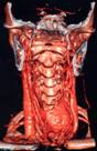
3D CT
ANGIOGRAPHY (CTA):
Full capabilities of
CTA including 3D
CTA RUN-OFF, Aorta, Carotid, Mesenteric, Renal Arteries and Coronary Arteries with
automatic Bolus Tracking technique
utilizing a sophisticated 3D/4D workstation.
·
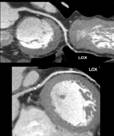
CORONARY
CALCIUM SCORING:
The most
sophisticated Coronary Calcium Scoring
software with Prospective
and Retrospective Cardiac Gating, automated scoring,
etc.
·
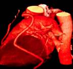
CORONARY
ANGIOGRAPHY:
The
3D Coronary CTA is an amazing and
spectacular new technology with sophisticated 3D/4D software allowing for
unfolding and flaying of the coronary arteries in 3D. Now
hard and soft plaques can be
visualized with automated software to determine the
degree of stenosis. Obviously,
CTA is done with a venous injection
rather than a femoral stick arterial catheterization. Any abnormality
requires a cardiology referral thus increasing referrals for cardiac cath.
This technique is nearly as accurate as conventional x-ray angiography.
·
CARDIAC
FUNCTIONAL ANALYSIS PACKAGE:
Functional
evaluation of the heart including Ejection
Fraction (EF), End Diastolic Volume (EDV), End Systolic Volume (ESV), Stroke
Volume (SV), Cardiac Output (CO), Wall Thickness, Wall Motion.
·
3D/4D
VIRTUAL COLONOSCOPY (VC):
Ultrafast VC with
real-time fly-through. Automatic multiplanar & 3D intraluminal display.
·
REAL-TIME CT FLUORO:
The only scanner
capable of real-time CT fluoroscopy.
This is the ultimate tool for performing real-time CT-guided procedures including
biopsies, aspirations, and arthrographic procedures in a matter
of a few minutes rather than 30-45 minutes. A
robot-like needle & syringe holder
allows for performing these procedures inside the gantry with
pinpoint
accuracy and
low dose without having to bringing
the patient out of the gantry.
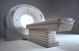
Advanced Imaging Center offers one of the world's most
sophisticated CT scanners. It is the only multi-slice, multi-detector
helical CT scanner in the Antelope Valley area. It is capable of
performing the most complex CT procedures.
CT (computed tomography) has been revolutionized by the
advent of VOLUMETRIC (spiral or helical) CT. In contrast to conventional
CT which images a slice of the body, incrementally moves the patient and
obtains another slice, etc., helical CT acquires data continuously as the
patient travels through the CT gantry.
The approach markedly decreases the time required to do
a study. The entire chest and abdomen are scanned in less than a minute.
Its other advantages are the elimination of motion
artifact, imaging during the window of optimal contrast enhancement for
maximal image contrast, and the ability to present images in any plane (multiplanar
reconstruction).
Advanced Imaging Center has installed the
state-of-the-art Picker (Marconi) Dual Slice Helical CT Scanner. This
spiral type of scanner is revolutionary in design. The radiation emitted
from the powerful x-ray tube is divided into two beams, and there are two
complete 360 degree sets of detectors. This concept allows scanning to
be performed very rapidly and with extremely high resolution. Slice
thickness of as little as .5 mm can be achieved. This is useful in
studying very small structures such as the bones in the middle ear.
Scanning of the chest, abdomen, and pelvis is performed
in a single breath-hold for each area. As an example, in scanning the
lungs, thin slices in the range of 2.5 to 5.0 mm can be obtained through
the entire chest during a single breath-hold, approximately 22 seconds.
The computer creates the images almost instantly and may be reviewed on
the monitor prior to the patient leaving the facility.
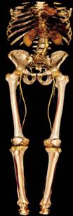
The scanner is also capable of performing
CT angiography,
imaging of the large arteries of the body with detail comparable to
traditional catheter angiography.
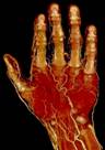
The scanner has many orthopedic applications because of
its powerful computer which allows information to be manipulated into
images in any plane. This information can be very helpful for surgical
planning. The computer has the ability to perform three-dimensional
surface images which may be rotated 360 degrees in any angle.
Oral
surgeons utilize the information obtained from scanning of the upper and
lower jaw for evaluation of bone detail and the location of nerves in
preparing for dental implants. The information provided cannot be
obtained from standard x-rays.
Q & A regarding AIC's Ultrafast, multidetector
Helical CT
Q. Can you tell me about the new helical CT
at AIC?
A. Certainly. It is a 16-slice CT. The new helical CT has
replaced our old dual-slice CT. It is a multi-slice (multi-detector) CT
capable of simultaneously scanning 16 slices evry 0.4 second (or 40 slices
per second), thus increasing the speed of CT scanning by at least a factor
of 40 or more. It is the fastest CT in the Antelope Valley area.
Q. What does ultrafast CT allow you to do?
A. Here's a summary (* denotes AIC exclusive):
- Fast high-resolution (1-2 mm) routine imaging of the neck,
chest, abdomen, and pelvis.
- *Fast high-res (0.5 mm) imaging of the IAC's/Temporal bones.
- *Fast ultra-high-resolution (0.5 mm) imaging of the bones for 4D
isotropic reconstruction.
- 3D and 4D CT Angiography (CTA): aorta, pulmonary arteries,
runoffs, brain.
- Coronary artery calcification scoring (excellent
non-invasive screening test).
- Virtual endoscopy (colonoscopy, bronchoscopy, and
endovascular angioscopy with CT!)
Q. What does coronary calcification scoring
tell you?
A. Coronary scoring is a safe, noninvasive and fast
screening CT technique that scans the heart in a few seconds and gives a
score based on the amount of calcium build up in the coronary arteries.
The score is a good predictor of future coronary events. For instance, it
has a 100% negative predictive value!
Q. Can you tell me more about the new
workstation at AIC?
A. Certainly. The new workstation is a state-of-the-art
silicon graphics workstation linked to all our modalities including
Open MRI, high-field MRI, Helical CT, and Nuclear med (SPECT/PET).
It is a powerful and sophisticated computer capable of amazing
multiplanar and 4D reconstruction, fusion of images from
different modalities, 4D virtual endoscopy, dental scan, 3D/4D CT
Angiography (CTA), just to name a few. It is simply technology at its
best.
Q. What is virtual endoscopy?
A. This noninvasive technique is one of the hottest areas in
CT today that allows 4D visualization of hollow organs (colon, bronchus,
etc.) similar to video endoscopy. This is only available at AIC.
Q. I have not heard of image fusion. Can you
explain?
A. Of course. Image fusion is a sophisticated software
utilized by our workstation that allows fusion of images from different
modalities (e.g., CT, MRI, SPECT, PET) or fusion of images from the same
modality at different times (to evaluate for growth). For example, a
physiologic/metabolic SPECT or PET image can be combined with an anatomic
CT or MRI image to provide an anatomico-physiologic image.
| ScanHealth |
Open MRI | High-field
MRI | MR Angiography | Helical CT |
CT Angiography |
Calcium Scoring |
| 4D CT Reconstruction |
Dental Scan | 4D
Ultrasound | Nuclear Medicine |
PET Scan |
DEXA Bone Density | X-ray |
Facts about services at AIC
 1.
The only community-based, private-practice, physician-operated
imaging facility in the Antelope Valley, just like any other private
practice medical office. Not belonging to any hospital or outside
imaging network. This means more personal and caring service.
1.
The only community-based, private-practice, physician-operated
imaging facility in the Antelope Valley, just like any other private
practice medical office. Not belonging to any hospital or outside
imaging network. This means more personal and caring service.
2. AIC was the first MRI-accredited site in the Antelope Valley
... approved by the American College of Radiology's MRI Accreditation
Committee.
3. Dr. Ray Hashemi is the only radiologist in the area with
fellowship training in ALL aspects of MRI, including neuro and
musculoskeletal MRI.
Why is AIC the PIONEER in advanced medical imaging in the
Antelope Valley?
1. AIC was the first to introduce a high-quality OPEN MRI
(open-air or open-sided MRI) to the Antelope Valley (January 1998).
2. AIC was the first to introduce Short-bore OPEN High-Field (1.5
Tesla) MRI to the Antelope Valley (January 1999).
3. AIC was the first to introduce multi-slice CT (MSCT) to the
Antelope Valley (August 1999); upgraded to a 16-slice CT in 2003.
4. AIC was the first to introduce revolutionary 3D Ultrasound to
the Antelope Valley (April 1999); upgraded to a GE 4D Ultrasound in
2004.
5. AIC was the first to introduce a PET scanner to the Antelope
Valley (July 1999).
6. AIC was the first to achieve MRI Accreditation in the Antelope
Valley (July 2000).
Call us at one of our
three locations: Lancaster (661) 949-8111, Palmdale (661) 456-2020
or
Valencia (661) 255-0060