MR Angiography is a noninvasive method to evaluate
the patency of blood vessels without resorting to invasive conventional
x-ray angiography. The latter requires insertion of a catheter into an
artery (usually in the groin) and carries a certain amount of risk
(infection, vessel rupture, hematoma, occlusion, embolism, allergies to
iodine dye, etc.)
Unlike x-ray angiogram, MR angiogram (MRA) is
performed either with no dye or with intravenous (as opposed to
intra-arterial) injection in an arm vein. The contrast or dye used in
MRA has no iodine in it and is much safer than x-ray dye.
MRA can be performed in the head, neck (carotids),
body, arm and legs.
At Advanced Imaging Center, MRA can be performed on
both the OPEN MRI and the high-field MRI. The latter has more MRA
applications, including contrast-enhanced MRA of the carotids, aorta,
pulmonary arteries, renal arteries, and extremities, not possible on an
open system as they required very high resolution and speed.
The high-field Siemens Symphony MRI system at AIC has
the most sophisticated MRA package available anywhere such as:
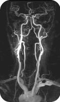
- 20-second Gadolinium-enhanced MRA of the carotids
utilizing an MR-compatible power injector;
- 30-second Gadolinium-enhanced body MRA (pulmonary
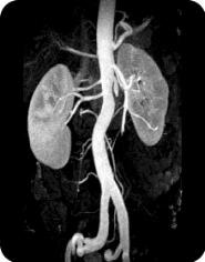
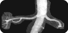
arteries, renal arteries, aorta, mesenteric angio;
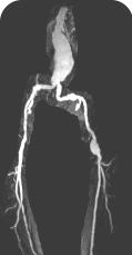
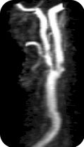
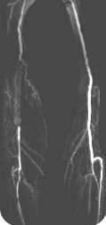
- 30-second Gadolinium-enhanced peripheral MRA (run-offs
utilizing a new automatic table-moving feature and a power injector
with accuracy similar to or better than X-ray angio).
- Exquisite ultra-resolution MRA of the circle of Willis and the
rest of the brain using Advanced MRA.
Contrast-Enhanced Subtraction MR Angiography
(CE-MRA): State-of-the-art
Q. What is Contrast-Enhanced MR Angiography (CE-MRA)?
CE-MRA is a fairly new, non-invasive MR
technique that utilizes ultra fast MR sequences to obtain exquisite
MRA images. These images are obtained during intravenous
administration of only 20-30 cc of Gadolinium using an MR-compatible
power injector. A bolus-tracking software tracks the bolus of
contrast for optimal timing. Subtraction techniques may also be
utilized for improved tissue contrast. Thus, a high-end scanner is
required to perform the procedure.
Q. What are some applications?
Carotids, brain, aorta, renals, pulmonary, run off, etc.
Q. How long does it take to do the carotids, for example?
The actual scan is performed in one breath hold
(about 20 seconds), but of course it takes some time to prepare the
patient and set up the protocols. The whole procedure can be performed
in ½ hour. 4D reconstruction is then performed.
Q. What is the accuracy of the test?
It is extremely accurate when performed properly,
and it correlates well with x-ray angiography and can replace it in
many instances. Some patients may undergo angioplasty or surgery based
on CE-MRA without the need for conventional angiography. 4D
reconstruction allows visualization at multiple angles. Best of all,
it is non-invasive!
 1.
The only community-based, private-practice, physician-operated
imaging facility in the Antelope Valley, just like any other private
practice medical office. Not belonging to any hospital or outside
imaging network. This means more personal and caring service.
1.
The only community-based, private-practice, physician-operated
imaging facility in the Antelope Valley, just like any other private
practice medical office. Not belonging to any hospital or outside
imaging network. This means more personal and caring service.